Where is the portal triad
By Avery Gonzales
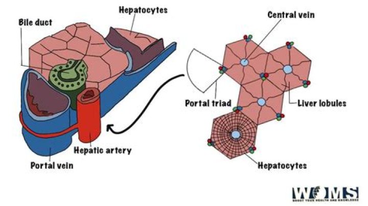
Portal areas (also called portal triads or portal canals) are located at the corners of liver lobules. Portal areas are normally surrounded by much larger areas packed with hepatic cords and sinusoids.
Where does the portal triad enter the liver?
Venous blood from the intestinal tract enters the liver through the portal triad via the portal vein, mixes with well-oxygenated hepatic arterial blood within the sinusoids, and exits via the terminal hepatic (central) vein.
How many portal triads are there?
Structure. The hepatic lobule can be described in terms of metabolic “zones”, describing the hepatic acinus (terminal acinus). Each zone is centered on the line connecting two portal triads and extends outwards to the two adjacent central veins.
What contains the portal triad?
The portal triad is contained within the hepatoduodenal ligament and contains the portal vein (posterolateral), hepatic artery (medial), and bile ducts (lateral) (figure 1). Variations in portal triad anatomy are relatively common (figure 2) [1].What makes up the portal triad ultrasound?
The “mickey mouse” sign is made up of the three portal structures. The left ear is the hepatic artery (HA), the right ear (at the arrow tip) is the common bile duct (CBD), and the portal vein is the face. Figure 7-5.
Does the portal vein drain into the IVC?
The portal venules drain into hepatic sinusoids that, in turn, are drained by the hepatic veins into the inferior vena cava.
Where is bile duct located?
The bile ducts are a series of thin tubes that go from the liver to the small intestine. Their main job is to allow a fluid called bile to go from the liver and gallbladder into the small intestine, where it helps digest the fats in food.
What do you mean by portal triad?
An anatomical unit of hepatic tissue, composed of an interlobular vein of liver, an interlobular artery of liver and an interlobular bile duct. The triads are embedded in the interlobular connective tissue and travel together throughout the liver parenchyma. ( NCI Thesaurus)Is hepatic vein and portal vein the same?
One is the hepatic artery, which brings in oxygen-rich blood from the heart. The other is the portal vein, which delivers blood from your stomach, intestines, and the rest of your digestive system.
What makes up the portal confluence?The portal vein is usually formed by the confluence of the superior mesenteric, splenic veins, inferior mesenteric, left, right gastric veins and the pancreatic vein. Conditions involving the portal vein cause considerable illness and death.
Article first time published onIs the portal vein Hepatopetal?
Because blood flow is normally hepatopetal in both the portal vein and the hepatic artery, opposite color signals in adjacent branches of these two circulations indicate hepatofugal portal vein flow. Figures 6. Hepatofugal flow in a patient with cirrhosis and portal hypertension.
What soft tissue structure houses the portal triad?
The porta hepatis transmits the portal triad—formed by the main portal vein, proper hepatic artery, and common hepatic duct—as well as nerves and lymphatics (1).
Where does the bile duct enter the duodenum?
The common bile duct passes through the pancreas before it empties into the first part of the small intestine (duodenum). The lower part of the common bile duct joins the pancreatic duct to form a channel called the ampulla of Vater or it may enter the duodenum directly.
Where does bile enter the digestive tract?
The common bile duct enters the small intestine at the sphincter of Oddi (a ring-shaped muscle), located a few inches below the stomach. About half the bile secreted between meals flows directly through the common bile duct into the small intestine.
How does bile enter the intestine?
The liver cells secrete the bile into small canals that lead to the common bile duct. From there, a smaller duct branches off and leads to the gallbladder. The common bile duct ends at the small intestine. The bile produced by the liver flows directly into the small intestine during a meal.
Does portal vein carry oxygenated or deoxygenated blood?
The liver receives a blood supply from two sources. The first is the hepatic artery which delivers oxygenated blood from the general circulation. The second is the hepatic portal vein delivering deoxygenated blood from the small intestine containing nutrients.
Where does the hepatic portal system begin and end?
The system extends from about the lower portion of the esophagus to the upper part of the anal canal. It also includes venous drainage from the spleen, pancreas and visceral fat.
What does the left portal vein branch into?
The main branches of the left portal vein originate from the umbilical portion, and supply liver segments 2, 3 and 4 5. The portal vein ramifies further, forming smaller venous branches and ultimately portal venules.
Where does the portal vein end?
The portal system drains the capillaries of the mesenteric and splenic veins and ends in the hepatic capillaries (Figure 76-1). The portal vein supplies partially oxygenated blood flow to the liver, supplementing the highly oxygenated blood flow of the hepatic artery to the liver.
Are portal veins Intersegmental vs Intersegmental?
The major branches of the portal veins run centrally within the segments (intrasegmental) with the exception of the ascending portion of the left portal vein, which runs in the left intersegmental fissure.
What is Hepatofugal flow in the main portal vein?
Hepatofugal flow (ie, flow directed away from the liver) is abnormal in any segment of the portal venous system and is more common than previously believed. Hepatofugal flow can be demonstrated at angiography, Doppler ultrasonography (US), magnetic resonance imaging, and computed tomography (CT).
What direction of flow is the portal vein?
When flow direction is normal in the portal vein (toward the liver), it is the same direction as the hepatic artery.
What is Portal area?
Abstract. The portal area is the ‘main entrance’ and one of the two main exits of the liver lobule. Through the main entrance portal and arterial blood reach the liver sinusoids. Through the exit the bile flows towards the duodenum.
What is Portal Hepatis?
Anatomical terminology The porta hepatis or transverse fissure of the liver is a short but deep fissure, about 5 cm long, extending transversely beneath the left portion of the right lobe of the liver, nearer its posterior surface than its anterior border.
What two ducts enter the duodenum?
The common bile duct and one pancreatic duct enter the duodenum via the major papilla.
Which ducts empty into the duodenum?
The common bile duct empties into the duodenum. The common bile duct is a tube-like structure that is formed where the common hepatic duct and the…
What ducts drains into the duodenum?
The common hepatic duct then joins with the cystic duct from the gallbladder to form the common bile duct. This runs from the liver to the duodenum (the first section of the small intestine).