What attaches to the calcaneus
By David Mccullough
The Achilles tendon is a tough band of fibrous tissue that connects the calf muscles to the heel bone (calcaneus). The Achilles tendon is also called the calcaneal tendon. The gastrocnemius and soleus muscles (calf muscles) unite into one band of tissue, which becomes the Achilles tendon at the low end of the calf.
Does the Achilles tendon attach to the calcaneal tuberosity?
The Achilles tendon connects the gastrocnemius and soleus muscles to the calcaneal tuberosity on the calcaneus (heel bone). The tendon begins near the middle of the calf, and receives muscle fibers on its inner surface, particularly from the soleus muscle, almost to its lower end.
What attaches to the posterior calcaneus?
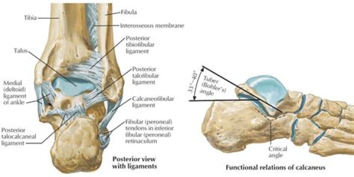
The posterior part of the calcaneus is circular, with three facets (superior, middle and inferior). The superior facet is separated from the calcaneal tendon by the retrocalcaneal bursa. The middle facet provides the attachment site for the calcaneal tendon (Achilles tendon).
What is calcaneal tuberosity?
cal·ca·ne·al tu·ber·os·i·ty. [TA] the posterior extremity of the calcaneus, or os calcis, forming the projection of the heel.Where does the calcaneus attach?
The calcaneus is the largest bone in the foot. It projects posterior to the tibia and fibula and acts as a short lever for the calf muscles (gastrocnemius and soleus) which insert onto its posterior surface via the Achilles tendon.
Where is the Achilles tendon attached?
The Achilles tendon is a strong fibrous cord that connects the muscles in the back of your calf to your heel bone.
Which muscle attaches to the femur and calcaneus?
OriginLateral head: Posterolateral aspect of lateral condyle of the femur Medial head: Posterior surface of medial femoral condyle, popliteal surface of femoral shaftInsertionPosterior surface of the calcaneus via the calcaneal tendonInnervationTibial nerve (S1, S2)
Where does Achilles tendon attach?
The Achilles tendon is a thick tendon located in the back of the leg. It connects the gastrocnemius and soleus muscles in the calf to an insertion point at the calcaneus (heel bone).Which muscle inserts to the calcaneus via the Achilles calcaneal tendon?
The soleus muscle, located deep/anterior to the medial and lateral gastrocnemius muscle heads, originates on the posterior aspect of the tibia (middle third of the medial border) and fibula (head and body) and inserts on the calcaneus through the Achilles tendon (see Figure 31.1).
What attaches to the Sustentaculum Tali?Several ligamentous structures attach to the sustentaculum tali: plantar calcaneonavicular ligament (anterior surface) deltoid ligament (medial surface) medial talocalcaneal ligament.
Article first time published onWhat is the quadratus plantae muscle?
Quadratus plantae makes part of the 20 individual foot muscles. It is situated in the second layer of muscles at the sole of the foot. The muscle consists of a lateral and medial head, coming together to form the bulk of this muscle.
Where does plantar fascia attach?
It involves inflammation of the plantar fascia — a tough, fibrous band of tissue that runs along the sole of the foot. The plantar fascia attaches to the heel bone (calcaneus) and to the base of the toes. It helps support the arch of the foot and has an important role in normal foot mechanics during walking.
What is inferior calcaneus?
An inferior calcaneal spur, also known as a plantar heel spur, is located on the lower aspect of the heel which is situated superior to the plantar fascia insertion. It develops as a response to plantar fasciitis over some time and may also be associated with ankylosing spondylitis especially in children.
Where is the calcaneal tuberosity?
The half of the bone closest to the heel is the calcaneal tuberosity. On its lower edge on either side are its lateral and medial processes (serving as the origins of the abductor hallucis and abductor digiti minimi).
What is attached to the heel bone?
The plantar fascia is a ligament attached to the heel bone (calcaneus). … These bones are called the metatarsal bones. Layers of muscles, tendons, nerves, and blood vessels run over the bottom of the foot.
Does calcaneus articulate with navicular?
The navicular bone is a keystone of the foot: it is part of the coxa pedis and articulates with the talus, first, second and third cuneiform, cuboid and calcaneus.
What muscle extends leg and stabilize knee two muscles?
The quadriceps femoris is one of the strongest muscle groups in the body that covers the anterior aspect of the femur. This group of muscles has a common function. They extend the leg at the knee joint. The rectus femoris has an additional role in stabilizing the hip joint and aiding in the flexion of the thigh.
Where does gastrocnemius attach?
gastrocnemius muscle, also called leg triceps, large posterior muscle of the calf of the leg. It originates at the back of the femur (thighbone) and patella (kneecap) and, joining the soleus (another muscle of the calf), is attached to the Achilles tendon at the heel.
What bones make up the tarsal?
The tarsal bones are 7 in number. They are named the calcaneus, talus, cuboid, navicular, and the medial, middle, and lateral cuneiforms.
Can you pull your Achilles?
The Achilles tendon can also tear or rupture, which might sound like a “pop” that seems to come from the back of your heel or calf. This needs immediate medical attention. Anyone can develop an Achilles tendon injury and it’s often linked to repetitive stress on the tendon.
Why is Achilles heel called that?
The term Achilles heel references a vulnerability or weakness. It is rooted in the myth of Achilles’ mother dipping him in the River Styx, making his entire body invulnerable except for the part of his foot where she held him—the proverbial Achilles heel. (Achilles tendon is an anatomical term.)
Can tight calf muscles cause Achilles pain?
Tight calf muscles can cause issues up and down ‘the chain’, downward tension would increase tension through your achilles tendon and onwards to the plantar fascia (in your feet). Upwards tension might give rise to issues in your hamstring’s or even your lower back.
Which muscle inserts at the calcaneus via the calcaneal tendon and is a weak knee flexor?
Origin and insertion The soleus muscle runs along the gastrocnemius muscle and together they insert onto the posterior surface of the calcaneus via the calcaneal tendon.
What two muscles attach distally to the calcaneal tendon quizlet?
What two muscles attach distally to the calcaneal tendon? These two muscles are known as the triceps surae and together are the most powerful plantar flexors of all the leg muscles.
Which of the following muscles attaches to the calcaneal bone through the calcaneal tendon quizlet?
What do the gastrocnemius and soleus muscles have in common? They both insert on the calcaneus via the calcaneal tendon.
Is the gastrocnemius a flexor or extensor?
The gastrocnemius is a biarticular muscle that acts not only as a plantar flexor, but also as a knee flexor, meaning that it is an antagonist during knee extension. In contrast, the soleus is a monoarticular plantar flexor.
What is the area above your heel called?
The Achilles tendon is the largest tendon in your body. It stretches from the bones of your heel to your calf muscles. You can feel it: a springy band of tissue at the back of your ankle and above your heel.
What ligaments are attached to the calcaneus?
Cervical ligament. Talocalcaneal interosseous ligament, Lateral, intermediate, and medial roots of the inferior extensor retinaculum. Bifurcate ligament.
Can the calcaneus be palpated?
The lateral wall of the calcaneus (E) can be palpated with little difficulty inferior and posterior to the tip of the fibula. If this lateral wall is palpated distal and inferior to the tip of the fibula, the peroneal tubercle (F) can be felt as the calcaneal neck nears the calcaneocuboid joint.
What is bifurcate ligament?
The bifurcated ligament (internal calcaneocuboid, interosseous ligament or bifurcate ligament) is a strong band, attached behind to the deep hollow on the upper surface of the calcaneus and dividing in front in a Y-shaped manner into a calcaneocuboid and a calcaneonavicular part.
What bone contains sustentaculum tali?
The sustentaculum tali is on the plantaromedial aspect of the calcaneus. Injuries involving the medial aspect of the hock often involve the sustentaculum tali and adjacent tarsal sheath.