
What are branchial arches
By Daniel Martin
The branchial arches are embryologic structures that develop into anatomic structures in the adult human. The term “branchial” derives from the Latin “branchia,” meaning gills, and is used to describe the development of many species of fish and amphibia.
What are the components of pharyngeal arches?
The components of each pharyngeal arch include an aortic arch, a specific cranial nerve and associated muscle, and a cartilage skeleton. The adult derivatives of each of these components are reviewed.
What structures are derived from the first pharyngeal arch?
The first pharyngeal arch–derived maxillary prominences fuse to form the intermaxillary segment which gives rise to the following oral cavity structures: philtrum of the lip, the maxilla and incisors, and the primary palate.
Are pharyngeal and aortic arches the same?
The aortic arches or pharyngeal arch arteries (previously referred to as branchial arches in human embryos) are a series of six paired embryological vascular structures which give rise to the great arteries of the neck and head. They are ventral to the dorsal aorta and arise from the aortic sac.What are pharyngeal pouches?
The pharyngeal pouches are endodermal-lined pockets that form on the INSIDE of the pharynx between the arches; pouch 1 forms between arch 1 and arch 2, pouch 2 forms between arch 2 and arch 3, etc. 1. Pharyngeal Pouch 1 –develops into the auditory tube and middle ear cavity. 2.
How many pharyngeal arches are there?
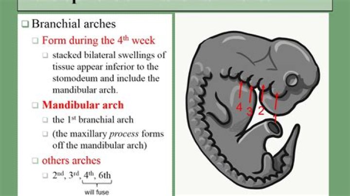
There are five pairs of pharyngeal arches in humans, and other amniotes, and these are numbered, from anterior to posterior, 1, 2, 3, 4 and 6 (Fig. 1). The 1st, most anterior, arch will form the jaws and the muscles of mastication, as well as the incus and malleus. This arch is innervated by the trigeminal nerve.
How many pharyngeal arches are present?
Pharyngeal arches, pouches, and clefts. There are five pairs of pharyngeal arches, numbered 1, 2, 3, 4, and 6 for comparative embryology reasons.
What are the 6 aortic arches?
ArchVessel4th Aortic sacR: Right subclavian artery L: Aortic arch Brachiocephalic artery (divides into right subclavian and right common carotid artery)5thR: Right pulmonary artery (proximal part) L: Ductus arteriosus6th Intersegmental arteryLeft pulmonary arteryWhat are the different branchial pouches?
Embryogenesis. The branchial apparatus consists of four pairs of arches separated externally by four paired grooves and internally by four paired pouches. The external grooves are called branchial clefts, and the internal pouches are known as pharyngeal pouches; they are separated by their branchial plates.
What are the different aortic arches?First aortic arch – regresses early, but a remnant forms a portion of the maxillary artery. … The left arch gives rise to the medial portion of the aortic arch. Fifth aortic arch – never forms or incompletely forms and regresses. Sixth aortic arch – The right and left arches separate into ventral and dorsal segments.
Article first time published onWhich organism develops breathing organs from pharyngeal?
In fishes and larvae of amphibians, these clefts develop gills and become respiratory organs. Pharyngeal pouches develop in the early embryos of all vertebrates, including the air-breathing terrestrial reptiles, birds, and mammals.
What branchial arches form the tongue?
The tongue begins to develop around the fourth week of intrauterine life. The first, second, third, and fourth pharyngeal arches contribute to the development of the various portions of the tongue. The development begins with the growth of a medial swelling from the first pharyngeal arch, known as tuberculum impar.
What pharyngeal arch the palatine tonsil is derived from?
The second pharyngeal pouch develops into the palatine tonsils, a secondary lymphoid organ playing a role in protecting the body from pathogens passing through the pharynx. The third pharyngeal pouch develops into the thymus and inferior portion of the parathyroid.
What is the pharyngeal apparatus?
The oral or pharyngeal apparatus serves the dual functions of respiration and feeding in many species in the animal kingdom. In humans, the mouth, nose and associated sinus, together with the pharynx which is connected to the larynx and trachea, form the upper respiratory tract.
Which pharyngeal arch makes the thyroid?
The thyroid initially arises caudal to the tuberculum impar, which is also known as the median tongue bud. This embryonic swelling develops from the first pharyngeal arch and occurs midline on the floor of the developing pharynx, eventually helping form the tongue as the two lateral lingual swellings overgrow it.
What are the symptoms of a pharyngeal pouch?
A pharyngeal pouch, also known as Zenker’s diverticulum, occurs when part of the pharyngeal lining herniates through the muscles of the pharyngeal wall. This occurs mainly in older people. Presenting symptoms include dysphagia, regurgitation of undigested food, halitosis, hoarseness, and chronic cough.
Where do we find pharyngeal pouches?
In the embryonic development of vertebrates, pharyngeal pouches form on the endodermal side between the pharyngeal arches. The pharyngeal grooves (or clefts) form the lateral ectodermal surface of the neck region to separate the arches. The pouches line up with the clefts, and these thin segments become gills in fish.
What are the 3 collateral branches of the aortic arch?
The aortic arch has three branches, the brachiocephalic trunk, left common carotid artery, and left subclavian artery. The aortic arch and its branches shown in situ. From its branches, the upper body, arms, head and neck.
Is aorta and aortic arch the same?
The aortic arch is the section of the aorta between the ascending and descending aorta. As it arises from the ascending aorta, the arch runs slightly backward and to the left of the trachea. … From this point on, it continues as the descending aorta.
What is Carotico systemic arch?
The left and right common carotid arteries supply the head and neck with oxygenated blood as they divide in the neck to form the external and internal carotid arteries. The aortic arch represents the continuation of the ascending aorta. The aorta originates from the left ventricle. So, the correct answer is option D.
What is a type 3 aortic arch?
If the origins of all the great vessels are included in the arc segment of the aortic arch subtended by the second index line, it is termed a type II arch. If the origins of all of the great vessels are included in the arc segment of the aortic arch subtended by the third index line, it is termed a type III arch.
Where is the proximal aortic arch?
The proximal thoracic aorta is the part of the aorta—the body’s largest artery—that runs through the chest.
Which arches disappear in all tetrapods?
- – No true internal gills→ so aortic arches do not.
- break up into afferent and efferent arteries.
- – I and II arches totally disappear in all tetrapods.
What animal will develop pharyngeal arches?
The pharyngeal arches, also known as visceral arches, are structures seen in the embryonic development of vertebrates that are recognisable precursors for many structures. In fish, the arches are known as the branchial arches, or gill arches.